Sunday, September 7, 2014

"Cross-section of a Tomato Stem" (Painting)

Titled "Cross-section of a Tomato Stem". The Painting measures at 8x10 inch on canvas. The Painting took two hours and was completed on September 6, 2014. I used only three colors, Turquoise, Black, and White--if you consider White as a color.

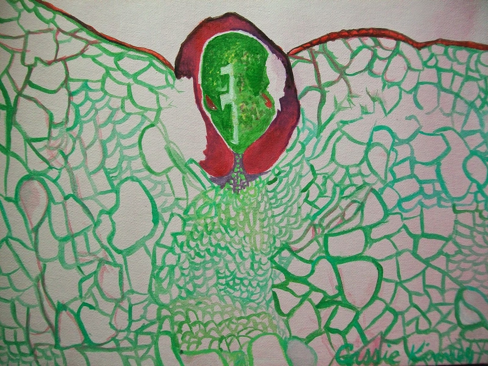
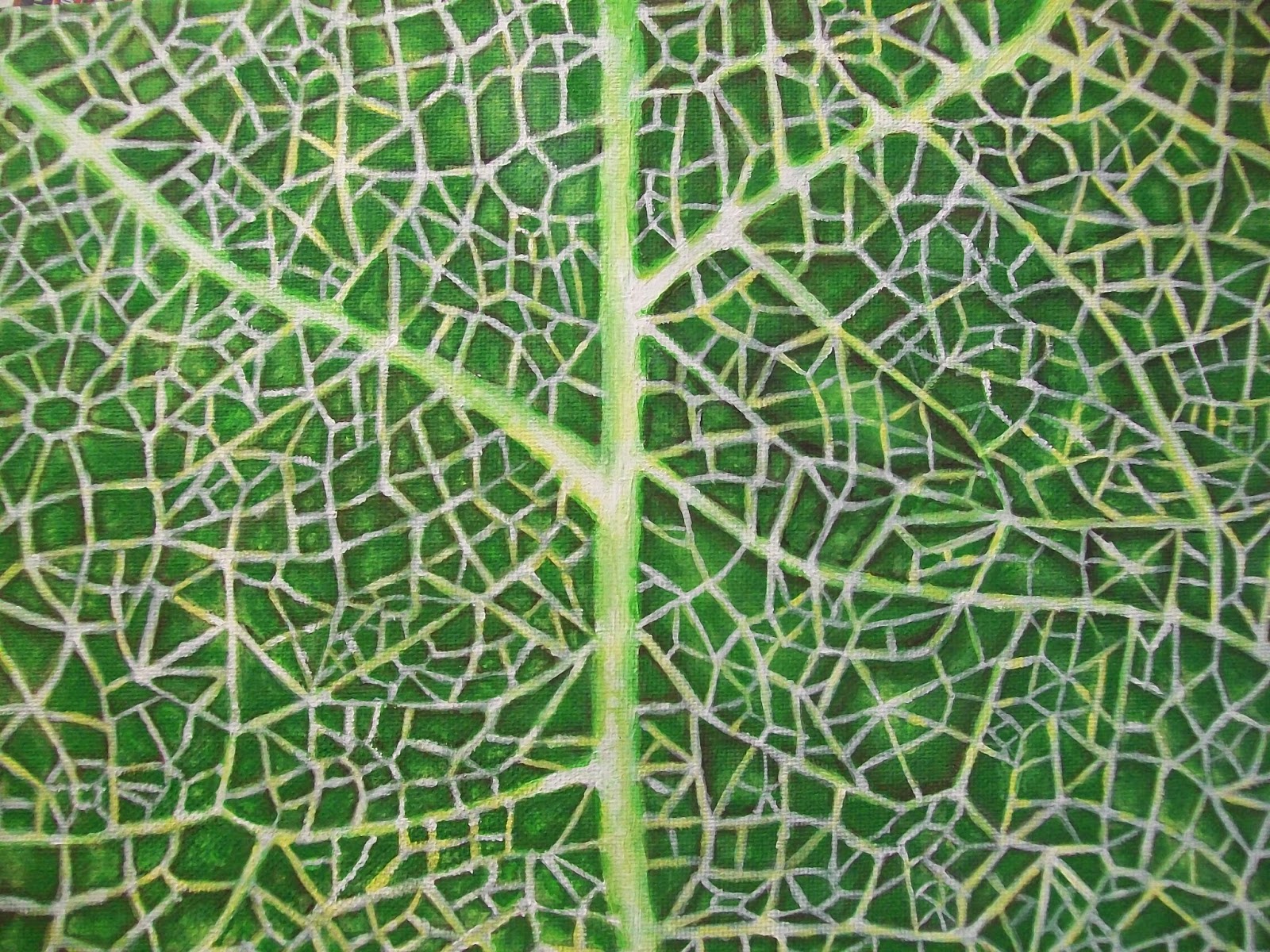
My last four paintings have been Animated microscopic images of plant tissues and their systems. See the other three in the links: "Strawberry (Fragaria) Embryo sporophyte with two cotyledons inside seed coat", "Microscope picture of Embryo development in Seed (Painting)", and "Veins of a leaf / Fractal Geometry".
Because I'm a gardener, I tend to be fascinated by the structures of plants. The Painting below is an Animated interpretation of a cross-section of a Tomato stem. The stems acts as an organ support, storage, and distribution of food and water. The cells you see have been divided over a period of time to develop the large Tomato plant. Certain cells, have different functions and altogether different appearance as you can see in the Painting below.
 For example, Xylem and Phloem are both complex, vascular tissues that thread through the ground tissue of the plant. But Xylem conducts water and dissolved mineral ions through vessel members while Phloem conducts food (sugars) through sieve tubes.
 I don't know if you have ever grown Tomatoes, but I have grown them as tall as six feet! Imagine the veins inside the stem which carry water from the roots to their leaves and flowers.


Saturday, September 6, 2014

"Heart-Shaped Embryo" (painting)

"Heart-Shaped Embryo and development of a seed, Shepherd's Purse (Capsella)"

The Biology textbook I have features many pictures of cells under a microscope. The pictures of cells under a microscope are like an extraordinarily intricate and colorful hallucination turned into an Abstract painting.
The painting I did here titled "Heart-Shaped Embryo and development of a seed, Shepherd's Purse (Capsella)" measures at 11x14 inch on canvas. The painting was translated from the Biology textbook by Starr, Evers, & Starr (seventh edition, page 460). As mentioned within the title, the painting captures the embryonic development of a seed from the Shepherd's Purse (Capsella) plant. The embryo is shaped much like a heart, the Basal cell below looks like a heart too, fixed on a spine. The "spine" is actually referred to as the "Suspender."
This painting was started on the 29th and I finished yesterday on August 30th, 2014. Immediately after finishing this painting (which only took 2 hours), I started and finished another Embryo painting yesterday, which you can see here: "Strawberry (Fragaria) Embryo sporophyte with two cotyledons inside seed coat". 


"Strawberry (Fragaria) Embryo"

"Strawberry (Fragaria) Embryo sporophyte with two cotyledons inside seed coat"


I mentioned in the last post my new fascination with observing cells under a microscope. Well, I've always been fascinated, but skimming through a Biology textbook led me to create two painting in the last two days that represented pictures of seeds under a microscope. In the last post, which you can see here "Microscope picture of Embryo development in Seed (Painting)", the embryo resembles a heart, but with this painting from the picture I referenced, the embryo looks like an 'Alien wearing a wig' (so my boyfriend says).
The painting below describes the Strawberry (Fragaria) where the seeds (ovaries) appear to the surface of the mature receptacle (Strawberry flesh).
This painting took (maybe) 30 minutes. I started and finished the painting on August 30th, 2014.

"Veins of a leaf / Fractal Geometry" (Painting)

The painting titled "Veins of a Leaf / Fractal Geometry" which measures 9x12 inches on canvas, was finished September 4th. The painting took approximately seven hours. I still may do finishing touches.
The idea behind this painting comes from the phenomenon that nature displays as branching of roots, or veins, or a mountain and rivers.

The reference photo is posted below painting.